
The Quiet Creep of Shock: Catching the Subtle Vital Sign Clues in Trauma Activations
Shock rarely announces itself with sirens. In the trauma bay, it often tiptoes in—polite, quiet, and dangerously convincing. Blood pressure looks “okay.” The patient is talking. The monitor hums along, unimpressed. And yet, somewhere between the triage vitals and the first CT slice, physiology is slipping. Early shock recognition is less about dramatic numbers and more about trends, context, and a healthy distrust of “normal.”
This article explores the subtle vital sign patterns that precede overt decompensation in trauma activations—and how clinicians can spot trouble before the crash cart earns its keep.
Why Early Shock Hides So Well
Compensatory mechanisms are excellent liars. Young, otherwise healthy trauma patients can maintain a normal systolic blood pressure despite significant hemorrhage through catecholamine-driven vasoconstriction and tachycardia (American College of Surgeons [ACS], 2018). By the time hypotension appears, the patient may already be deep into oxygen debt.
Relying on single-point vital signs is like judging a movie from one frame. Shock is a trajectory, not a snapshot.
Subtle Vital Sign Trends That Matter More Than “Normal”
1. Heart Rate: It’s the Direction, Not the Destination
A heart rate of 98 bpm may not raise eyebrows—but a progression from 72 → 86 → 98 in 20 minutes should. Persistent or rising tachycardia, especially when pain and anxiety are addressed, is an early marker of hypovolemia (ACS, 2018).
Even more telling is the Shock Index (SI)—heart rate divided by systolic blood pressure. An SI ≥ 0.9 is associated with increased mortality and transfusion needs, even when absolute blood pressure is normal (Rady et al., 1994; Mutschler et al., 2013).
Clinical pearl: The patient doesn’t need to be hypotensive to be in trouble—your math already told you.
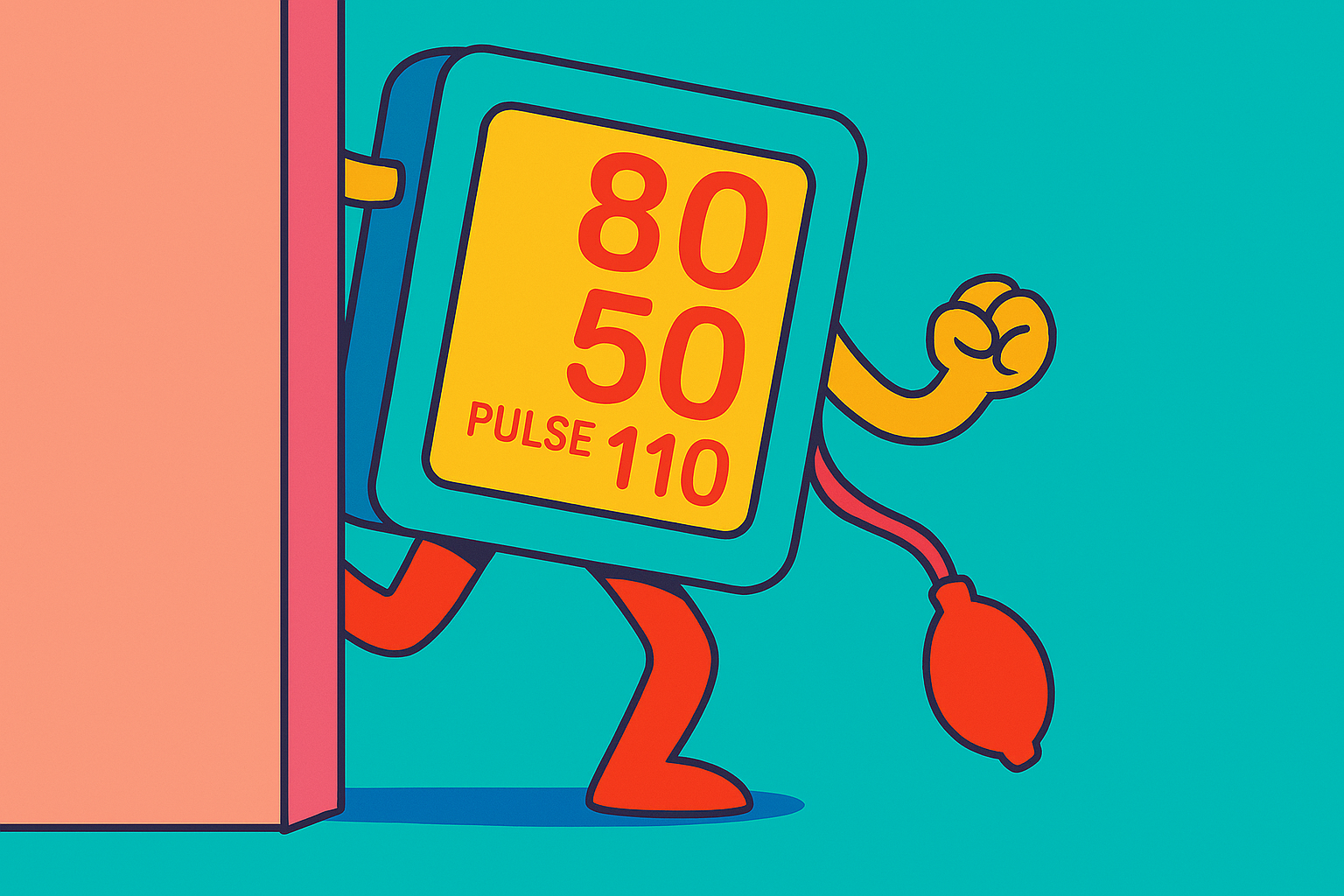
2. Blood Pressure: The False Reassurance of “120/80”
Systolic blood pressure is a late marker of hemorrhagic shock. Early on, you may see:
Narrowing pulse pressure, reflecting reduced preload and stroke volume
A “stable” SBP that requires increasing sympathetic tone to maintain
Permissive hypotension principles remind us that “normal” pressures are not always the goal—and certainly not proof of adequate perfusion (Cannon et al., 2017).
3. Respiratory Rate: The Most Ignored Red Flag
Respiratory rate is both under-measured and under-appreciated. A creeping rise—18 → 22 → 26—may reflect metabolic acidosis compensation well before labs return. Multiple studies identify respiratory rate as one of the earliest vital sign changes in shock states, often preceding hypotension (ACS, 2018).
If you trust only one vital sign, make it this one.
4. End-Tidal CO₂: The Perfusion Canary
In intubated or sedated trauma patients, declining end-tidal CO₂ (EtCO₂) can signal reduced cardiac output and worsening perfusion—even with unchanged ventilator settings. Low EtCO₂ values have been associated with increased mortality in trauma and hemorrhagic shock (Helm et al., 2002).
Think of EtCO₂ as a real-time report card for circulation, not just ventilation.
5. Mental Status and Skin: Old School, Still Gold
Subtle agitation, delayed responses, or a shift from calm to restless can reflect cerebral hypoperfusion. Likewise, cool, clammy skin and delayed capillary refill remain valuable bedside clues when interpreted in context (ACS, 2018).
Technology is helpful—but your eyes and hands still matter.
Laboratory Trends That Confirm the Suspicion
Lactate and Clearance
An elevated lactate indicates global hypoperfusion, but failure to clear lactate over time is an even stronger predictor of mortality (Abramson et al., 1993). A single value is informative; the trend is decisive.
Base Deficit
Base deficit correlates with the severity of shock and transfusion requirements. Worsening base deficit, even with stable vitals, suggests ongoing oxygen debt (Davis et al., 1996).
Putting It Together: Think Like a Trend Detective
Early shock recognition isn’t about finding the abnormal number—it’s about connecting small deviations across systems:
Rising HR + stable BP
Increasing RR + subtle mental status change
Normal SBP + elevated shock index
“Okay” vitals + worsening lactate
When these clues cluster, believe them.
Call to Action: Make Trends the Trauma Team’s Shared Language
Early shock recognition saves lives—but only if teams act on it. Here’s the challenge:
Trend vital signs out loud during trauma activations
Incorporate shock index and lactate trends into routine handoffs
Empower nurses and trauma narrators to voice concern when patterns feel wrong—even if numbers look “fine”
Teach new clinicians that normal vitals do not equal normal physiology
Shock whispers before it screams. Our job is to listen sooner.
References
Abramson, D., Scalea, T. M., Hitchcock, R., Trooskin, S. Z., Henry, S. M., & Greenspan, J. (1993). Lactate clearance and survival following injury. The Journal of Trauma, 35(4), 584–589. https://pubmed.ncbi.nlm.nih.gov/8411283/
American College of Surgeons. (2018). ATLS®: Advanced Trauma Life Support® student course manual (10th ed.). https://www.facs.org/quality-programs/trauma/education/atls/
Cannon, J. W., Khan, M. A., Raja, A. S., Cohen, M. J., Como, J. J., Cotton, B. A., … Neal, M. D. (2017). Damage control resuscitation in patients with severe traumatic hemorrhage. Journal of Trauma and Acute Care Surgery, 83(3), 605-617. https://doi.org/10.1097/TA.0000000000001333
Davis, J. W., Parks, S. N., Kaups, K. L., Gladen, H. E., & O’Donnell-Nicol, S. (1996). Admission base deficit predicts transfusion requirements and risk of complications. The Journal of Trauma, 41(5), 769–774. https://doi.org/10.1097/00005373-199611000-00001
Helm, M., Hauke, J., & Lampl, L. (2002). A prospective study of the quality of pre-hospital emergency ventilation in patients with severe head injury. British Journal of Anaesthesia, 88(3), 345–349. https://doi.org/10.1093/bja/88.3.345
Mutschler, M., Nienaber, U., Münzberg, M., Wölfl, C., Schoechl, H., Paffrath, T., … Maegele, M. (2013). The Shock Index revisited—a fast guide to transfusion requirement? Critical Care, 17(4), R172. https://doi.org/10.1186/cc12851
Rady, M. Y., Smithline, H. A., Blake, H., Nowak, R., & Rivers, E. (1994). A comparison of the shock index and conventional vital signs to identify acute, critical illness in the emergency department. Annals of Emergency Medicine, 24(4), 685–690. https://doi.org/10.1016/S0196-0644(94)70279-9